
A microscope is a special tool that helps us see very tiny things that our eyes cannot see normally. Imagine trying to see a single ant’s hair—impossible! A microscope makes it bigger and clearer. Scientists use it to look at germs, blood cells, and insects up close. It works using glass lenses that bend light to zoom in, just like a magnifying glass, but much more powerfully!
Types of Microscopes
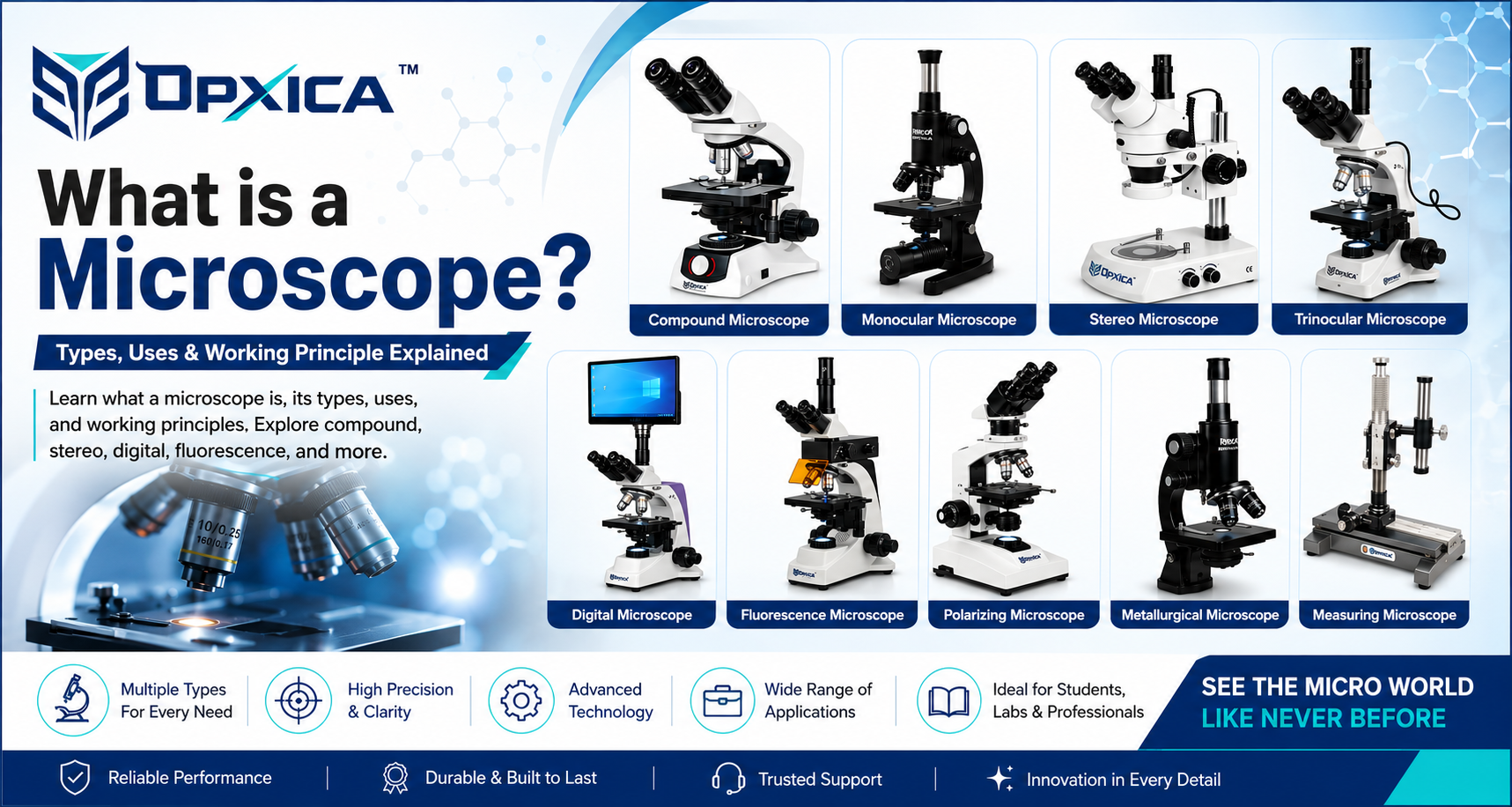
Microscopes come in different types, each designed for a specific purpose. Let’s explore the most common types in a simple and easy way!
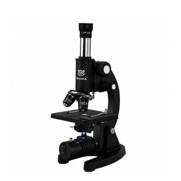
1. Light Microscope (Optical Microscope)
This is the most common and basic type of microscope. It uses visible light and glass lenses to magnify objects. You will find this microscope in almost every school and college laboratory.
It can magnify objects up to 100 times their actual size. Scientists use it to observe blood cells, plant cells, bacteria, and small insects. It is simple to use, affordable, and perfect for beginners. When you think of a microscope, this is the one that comes to mind first!
Uses of a Light Microscope
- Used to observe and analyze microorganisms, cells, and tissues in laboratories.
- Helps in studying plant and animal cell structures for biological research.
- Widely used in medical laboratories for blood sample and tissue examination.
- Assists educational institutions in teaching biology and life science subjects.
- Supports microbiological and clinical research by enabling detailed specimen observation.
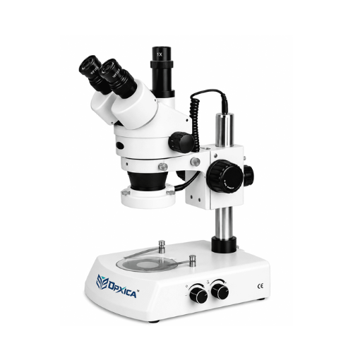
2. Stereo Microscope (Dissecting Microscope)
This microscope is used to look at larger objects in 3D. Unlike other microscopes, it does not magnify too much — only about 7 to 45 times. But it gives a three-dimensional view, which makes it very useful.
Jewelers use it to work on small ornaments. Scientists use it to dissect insects, rocks, coins, and plants. It is also used in surgery to perform delicate operations. It is easy to use and very helpful in detailed hand work.
Uses of a Stereo Microscope (Dissecting Microscope)
- Used for examining the surface details of insects, plants, electronic components, and small objects.
- Helps in dissection procedures by providing a clear three-dimensional view of specimens.
- Widely used in quality control and inspection of manufactured products.
- Assists jewelers, watchmakers, and technicians in handling and repairing delicate items.
- Supports educational and research laboratories in studying larger specimens at low magnification.
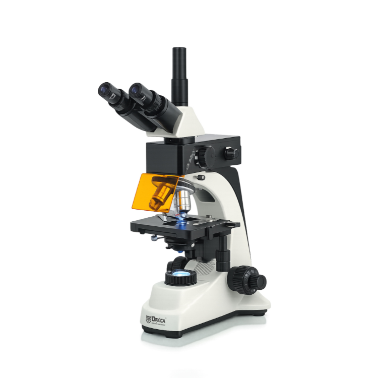
3. Fluorescence Microscope
This is a special type of microscope that uses ultraviolet (UV) light to make certain objects glow in the dark. Special dyes called fluorescent dyes are applied to the specimen, and when UV light hits them, they light up in bright colors.
Scientists use it to study cancer cells, bacteria, and proteins inside living cells. It is mostly used in medical research and hospitals to detect diseases at an early stage.
Uses of a Fluorescence Microscope
- Used to detect and study specific cells, proteins, and microorganisms using fluorescent dyes.
- Helps in medical diagnostics for identifying infectious diseases and cellular abnormalities.
- Widely used in molecular biology and biotechnology research.
- Assists researchers in observing cellular structures and biological processes in detail.
- Supports pharmaceutical and clinical laboratories in drug development and disease studies.
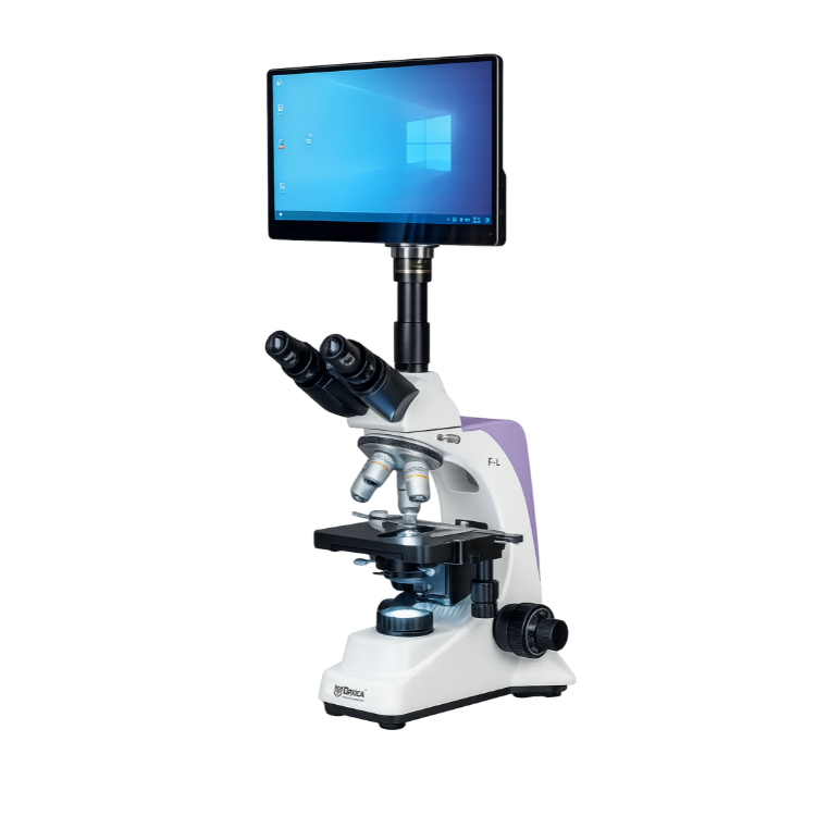
4. Digital Microscope
This is a modern and smart microscope that connects to a computer or smartphone. It uses a digital camera to capture and display magnified images on a screen. You can save, share, and analyze images easily.
It is very user-friendly and perfect for students, teachers, and beginners. It is widely used in education, quality checking in factories, and even at home for fun experiments.
Uses of a Digital Microscope
- Used for capturing, viewing, and analyzing high-resolution images directly on a computer or display screen.
- Helps in educational and research laboratories for detailed specimen observation and documentation.
- Widely used in electronics and manufacturing industries for component inspection and quality control.
- Assists forensic experts in examining evidence, fibers, and other microscopic materials.
- Supports medical, biological, and industrial applications by enabling image storage, measurement, and sharing.

5. Polarizing Microscope
This is a special microscope that uses polarized light to study certain materials. Normal light vibrates in all directions, but polarized light vibrates in only one direction. This makes it easier to study materials that react differently to light.
It is widely used by geologists to study rocks and minerals, as different minerals show beautiful colors and patterns under polarized light. It is also used in the chemical and pharmaceutical industry to study crystals and chemical compounds. It is a very important tool in material science and geology studies.
Uses of a Polarizing Microscope
- Used to identify and analyze minerals, rocks, and geological specimens.
- Helps in studying crystals and chemical compounds in pharmaceutical and chemical laboratories.
- Widely used in material science for examining the optical properties of materials.
- Assists researchers in detecting stress, defects, and structural variations in transparent materials.
- Supports forensic, industrial, and academic research applications requiring polarized light analysis.

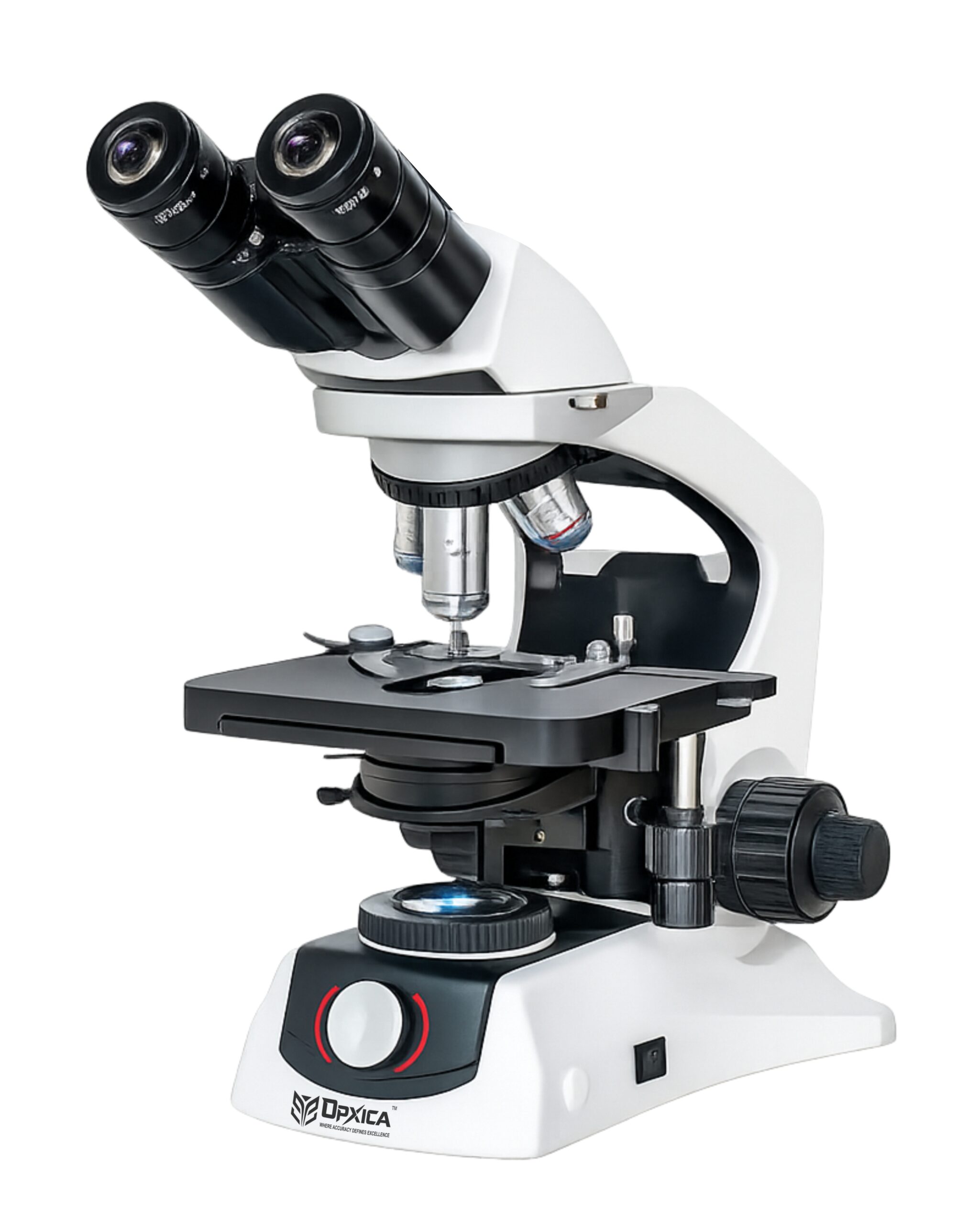
6. Compound Microscope
This is one of the most popular microscopes used in schools, colleges, and laboratories. It is called a compound microscope because it uses two sets of lenses — the eyepiece lens and the objective lens — working together to magnify the object.
It can magnify objects from 40 times to 100 times their actual size. It is used to observe bacteria, fungi, blood cells, and tiny organisms. The compound microscope is perfect for students because it is easy to handle, affordable, and gives clear images. It is the most commonly used microscope in everyday science education.
Uses of a Compound Microscope
- Used to observe bacteria, fungi, blood cells, and other microscopic organisms.
- Helps students and researchers study cell structures and biological specimens in detail.
- Widely used in schools, colleges, and laboratories for science education and practical experiments.
- Assists medical laboratories in examining blood samples, tissues, and microorganisms.
- Supports microbiology, pathology, and biological research by providing high magnification and clear imaging.
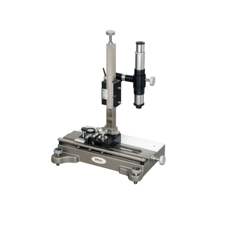
7. Travelling Microscope
This is a unique and special type of microscope that is used to make very accurate measurements of small objects and distances. Unlike other microscopes that are fixed in one place, this microscope can travel or move horizontally and vertically on a precise scale.
It is mostly used in physics laboratories to measure the diameter of a thin wire, capillary tube, or the wavelength of light. It has a vernier scale attached to it for extremely accurate readings. It is not used to study living things but is a very important tool for precise scientific measurements in experiments.
Uses of a Travelling Microscope
- Used for precise measurement of small distances, diameters, and dimensions in physics experiments.
- Helps in measuring the diameter of thin wires, capillary tubes, and small objects with high accuracy.
- Widely used in optical experiments for determining the wavelength of light and refractive index.
- Assists researchers and students in conducting accurate laboratory measurements using a vernier scale.
- Supports scientific and engineering applications where precise linear measurements are required.
How Different Types of Microscopes Work
1. Light Microscope (Optical Microscope)
A light microscope works by shining visible light through or onto a specimen. The light passes through glass lenses called the objective lens and eyepiece lens. These lenses bend and focus the light to create a magnified image that appears larger to the viewer.
Working Principle:
- Light illuminates the specimen.
- Objective lens magnifies the image.
- Eyepiece lens further enlarges the image.
- The user sees a magnified view of the specimen.
2. Stereo Microscope (Dissecting Microscope)
A stereo microscope uses two separate optical paths and two objective lenses to create a three-dimensional (3D) image. Instead of viewing a flat image, each eye sees the object from a slightly different angle.
Working Principle:
- Light reflects from the surface of the object.
- Two separate optical systems capture the image.
- The brain combines both images.
- A realistic 3D view is produced.
3. Fluorescence Microscope
A fluorescence microscope uses ultraviolet (UV) or blue light to excite fluorescent dyes attached to the specimen. These dyes absorb the light and emit bright-colored light, making specific structures visible.
Working Principle:
- Fluorescent dyes are applied to the specimen.
- UV light excites the dyes.
- The dyes emit colored light.
- Special filters capture the emitted light and create an image.
4. Digital Microscope
A digital microscope replaces the traditional eyepiece with a digital camera. The camera captures the magnified image and displays it on a computer, tablet, or monitor.
Working Principle:
- Light illuminates the specimen.
- Lenses magnify the image.
- A digital sensor captures the image.
- The image is displayed on a screen for viewing and analysis.
5. Polarizing Microscope
A polarizing microscope uses polarized light instead of ordinary light. Polarizing filters allow light to vibrate in only one direction, helping scientists observe materials that interact with light differently.
Working Principle:
- Light passes through a polarizer.
- Polarized light enters the specimen.
- The specimen changes the light’s direction and intensity.
- An analyzer filter reveals colors and patterns for study.
6. Compound Microscope
A compound microscope works using two lens systems: the objective lens and the eyepiece lens. The objective lens creates an enlarged image, and the eyepiece magnifies it further.
Working Principle:
- Light passes through the specimen.
- Objective lens creates a magnified image.
- Eyepiece lens enlarges the image again.
- The viewer observes a highly magnified specimen.
7. Travelling Microscope
A travelling microscope combines optical magnification with a precision measuring scale. The microscope can move horizontally and vertically along calibrated scales.
Working Principle:
- The specimen is viewed through the microscope.
- The microscope is moved precisely using adjustment screws.
- Vernier scales measure the displacement.
- Accurate dimensions and distances are calculated.
Quick Summary Table
| Type | Uses Light? | Magnification | Best For |
|---|---|---|---|
| Light Microscope | ✅ Yes | Up to 100x | Students, basic science |
| Electron Microscope | ❌ No (Electrons) | Up to 1,000,000x | Advanced research |
| Stereo Microscope | ✅ Yes | 7x to 45x | 3D viewing, surgery |
| Fluorescence Microscope | UV Light | High | Medical research |
| Digital Microscope | ✅ Yes | Varies | Students, digital use |
| Polarizing Microscope | ✅ Polarized Light | Up to 100x | Geology, minerals, crystals |
| Compound Microscope | ✅ Yes | 40x to 100x | Schools, colleges, labs |
| Travelling Microscope | ✅ Yes | Moderate | Physics measurements |
Conclusion
Although all microscopes are designed to magnify objects, each type uses a different technology. Light and compound microscopes rely on glass lenses and visible light, fluorescence microscopes use UV light and dyes, digital microscopes use cameras, polarizing microscopes use polarized light, stereo microscopes provide 3D viewing, and travelling microscopes focus on precise measurements. Together, they help scientists, students, researchers, and engineers explore details that are invisible to the naked eye.