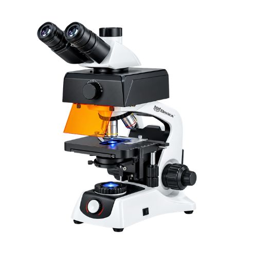
Description
Technical Specifications
| Parameter | Specification |
|---|---|
| Product Name | Fluorescent Microscope OPX-257 |
| Model Number | OPX-257 |
| Microscope Type | Research Fluorescent Microscope |
| Optical System | Infinity-Corrected Optical System |
| Viewing Head | Trinocular Head |
| Head Inclination | 30° Inclined |
| Interpupillary Distance | 48–75 mm |
| Eyepieces | High Wide-Field 10X Extra-Wide-Field Eyepieces |
| Objectives | Plan-Achromatic Objectives |
| Objective Range | 4X to 100X Oil Immersion |
| 4X Objective | NA 0.10 |
| 100X Objective | Oil Immersion, NA 1.25 |
| Nosepiece | Inward-Facing Quadruple Revolving Nosepiece |
| Stage Type | Double-Layer Mechanical Stage |
| Stage Size | 210 × 40 mm |
| Stage Movement | Smooth X-Y Movement |
| Condenser | Rack & Pinion Condenser |
| Condenser NA | 1.25 |
| Condenser Adjustment | Adjustable Light-Path Alignment |
| Fluorescence Illumination | 100W HBO Ultra-Voltage Mercury Lamp |
| Fluorescence Brightness | Adjustable |
| Bright-Field Illumination | Available |
| Fluorescence Filters | Multi-Channel Blue, Green & UV Filters |
| Blue Filter Range | 420–485 nm |
| Green Filter Range | 480–550 nm |
| UV Filter Range | 330–400 nm |
| Magnification Range | 40X–2500X |
| Maximum Magnification | Up to 2500X with Oil Immersion |
| Applications | Immunofluorescence, Cell Biology, Pathology, Fluorescent Staining, Venereal Disease Testing |
| Brand | OPXICA |
Key Features
Infinity-Corrected Optical System
The OPX-257 uses an infinity-corrected optical system designed to provide clear, high-resolution images and support advanced microscopy applications.
Trinocular Observation Head
The 30° inclined trinocular head provides comfortable specimen observation while allowing a camera to be connected for digital imaging and documentation.
Plan-Achromatic Objectives
The microscope incorporates plan-achromatic objectives ranging from 4X to 100X oil immersion, providing broad magnification capability and improved image correction across the viewing field.
Up to 2500X Magnification
With the appropriate oil-immersion objective configuration, the OPX-257 provides magnification up to 2500X, making it suitable for high-magnification research and diagnostic observation.
Multi-Channel Fluorescence System
The microscope supports multiple fluorescence wavelength ranges:
- Blue: 420–485 nm
- Green: 480–550 nm
- UV: 330–400 nm
This multi-channel configuration supports different fluorescence-based observation requirements.
100W HBO Mercury Lamp
A 100W HBO ultra-voltage mercury lamp provides fluorescence excitation. Brightness can be adjusted according to the observation requirements.
Bright-Field and Fluorescence Observation
The OPX-257 combines conventional bright-field microscopy with fluorescence microscopy in a single system, allowing users to select the appropriate observation method for their specimen.
Precision Mechanical Stage
The double-layer mechanical stage provides smooth X-Y specimen movement and controlled positioning during microscopic examination.
Rack & Pinion Condenser
The rack-and-pinion condenser with NA 1.25 provides controlled illumination and adjustable light-path alignment for optimized specimen observation.
Working Purpose
The Fluorescent Microscope OPX-257 is designed for advanced observation of biological specimens using both conventional bright-field and fluorescence microscopy techniques.
It is suitable for:
- Immunofluorescence research
- Cell biology
- Pathology research
- Fluorescent staining
- Microbiological studies
- Cellular morphology
- Biological specimen examination
- Diagnostic laboratory research
- Venereal disease testing
- Pharmaceutical research
- Biotechnology research
- Academic and scientific research
Applications
Immunofluorescence Research
The multi-channel fluorescence system can be used for research involving fluorescently labelled antibodies, cells, tissues, and other biological specimens.
Cell Biology
Suitable for studying cellular structures, morphology, fluorescent markers, and biological processes.
Pathology Laboratories
Can be used for research and advanced examination of stained tissue and cellular specimens.
Microbiology
Useful for fluorescence-based observation and identification workflows involving suitable labelled microorganisms and biological samples.
Pharmaceutical & Biotechnology Research
Suitable for laboratory research involving cells, tissues, fluorescent markers, and biological assays.
Medical & Research Institutions
Designed for advanced microscopy applications in universities, research institutes, diagnostic research centers, and specialized laboratories.
Highlight Points
- High-end research and diagnostic applications
- Infinity-corrected optical system
- Trinocular 30° inclined viewing head
- 48–75 mm interpupillary adjustment
- Plan-achromatic objectives
- 4X to 100X oil-immersion objective range
- Up to 2500X magnification
- Multi-channel fluorescence capability
- Blue, Green and UV fluorescence filters
- 100W HBO mercury fluorescence lamp
- Adjustable fluorescence brightness
- Bright-field observation capability
- Double-layer mechanical stage
- Smooth X-Y specimen movement
- NA 1.25 rack-and-pinion condenser
- Suitable for immunofluorescence and cell biology research
Advantages of OPX-257
The OPX-257 provides a combined platform for bright-field and fluorescence microscopy, reducing the need for separate microscope systems for different observation techniques. Its trinocular configuration also supports digital imaging and documentation when paired with a compatible microscope camera.
The combination of plan-achromatic optics, multi-channel fluorescence filters, high-intensity mercury-lamp excitation, precision mechanical movement, and oil-immersion magnification makes the microscope suitable for advanced laboratory and research environments.
Frequently Asked Questions (FAQs)
1. What is a fluorescent microscope?
A fluorescent microscope is designed to observe specimens that emit fluorescence after being illuminated with specific excitation wavelengths. It is commonly used in cell biology, immunofluorescence, pathology, microbiology, biotechnology, and scientific research.
2. What is the magnification range of the OPX-257?
The OPX-257 provides a magnification range of 40X to 2500X, with the maximum magnification achieved using the appropriate oil-immersion objective configuration.
3. What type of objectives does the OPX-257 use?
The microscope uses plan-achromatic objectives from 4X through 100X oil immersion. The 100X oil objective has a numerical aperture of 1.25.
4. What fluorescence filters are available?
The OPX-257 supports three primary fluorescence wavelength ranges:
- Blue: 420–485 nm
- Green: 480–550 nm
- UV: 330–400 nm
5. What light source is used for fluorescence?
The OPX-257 uses a 100W HBO ultra-voltage mercury lamp for fluorescence excitation, with adjustable brightness.
6. Can the OPX-257 perform bright-field microscopy?
Yes. The microscope supports bright-field observation in addition to fluorescence microscopy, allowing conventional and fluorescence-based examination on the same platform.
7. Can a camera be connected to the OPX-257?
Yes. The trinocular head provides a dedicated optical path for connecting a compatible microscope camera for digital imaging, recording, and documentation.
8. What is the purpose of the 100X oil-immersion objective?
Oil immersion is used with high numerical-aperture objectives to improve optical resolution and light collection. The 100X oil-immersion objective is useful for detailed examination of appropriately prepared specimens.
9. What is immunofluorescence microscopy?
Immunofluorescence microscopy is a technique that uses fluorescently labelled antibodies or related probes to visualize specific biological targets. The OPX-257’s multi-channel fluorescence configuration can support suitable immunofluorescence research applications.
10. Is the OPX-257 suitable for cell biology research?
Yes. Its fluorescence illumination, multi-channel filters, plan-achromatic objectives, and high magnification make it suitable for various cell biology research applications.
11. Is the OPX-257 suitable for pathology?
The microscope can be used for pathology research, fluorescent staining, tissue examination, and related laboratory applications. Specific diagnostic use should be based on the applicable laboratory procedures, accessories, and regulatory requirements.
12. What is the stage size?
The OPX-257 uses a double-layer mechanical stage measuring 210 × 40 mm, providing controlled X-Y specimen movement.
13. What type of condenser is used?
The microscope uses a rack-and-pinion condenser with NA 1.25 and adjustable light-path alignment.
14. Who can use the OPX-257?
The microscope is suitable for:
- Research laboratories
- Biotechnology laboratories
- Pathology research centers
- Medical research institutions
- Pharmaceutical laboratories
- Universities
- Cell biology laboratories
- Microbiology laboratories
- Scientific research institutes
15. How should the OPX-257 be maintained?
For reliable operation:
- Clean optical components using appropriate lens-cleaning materials.
- Avoid touching optical surfaces with bare fingers.
- Keep the microscope covered when not in use.
- Keep the mechanical stage clean.
- Follow appropriate procedures when handling the mercury lamp.
- Allow the illumination system to operate and cool according to the manufacturer’s instructions.
- Store the microscope in a clean and dry environment.
- Handle fluorescence filters and optical components carefully.
16. Is the OPX-257 available for bulk orders?
Yes. OPXICA can supply the Fluorescent Microscope OPX-257 for institutional, research, laboratory, distributor, and bulk requirements. Accessories, camera systems, and configuration requirements can also be discussed with OPXICA.
17. Where can I buy the OPXICA Fluorescent Microscope OPX-257?
The OPXICA Fluorescent Microscope OPX-257 can be purchased directly from OPXICA. Contact OPXICA for current pricing, availability, institutional quotations, accessories, camera options, and technical support.