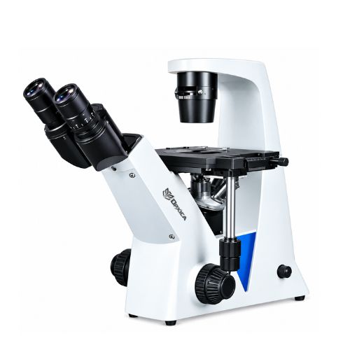
Description
Technical Specifications
| Parameter | Specification |
|---|---|
| Product Name | Inverted Phase Contrast Tissue Culture Microscope OPX-176 |
| Brand | OPXICA |
| Model No. | OPX-176 |
| Microscope Type | Inverted Phase Contrast Tissue Culture Microscope |
| Standard Function | Bright Field (BF) + Phase Contrast (PH) |
| Optical System | Infinity Plan |
| Optical Configuration | Infinity Plan & Phase Contrast |
| Standard Magnification | 100X–400X |
| Viewing Head | Seidentopf Trinocular |
| Head Inclination | 45° |
| Head Rotation | Eyetube Rotates 360° |
| Interpupillary Distance | 48–76 mm |
| Head Diopter | Both Tubes Adjustable |
| Light Path | E100:P0 / E20:P80 |
| Eyepieces | WF10X/22 mm, 2 pcs |
| Eyepiece Diameter | 30.0 mm |
| Eyepiece Adjustment | Diopter Adjustable |
| Nosepiece | 5-Hole Revolving Nosepiece |
| Objectives | Infinity Plan LWD Semi-APO FL 10X, 20X, 40X |
| Phase Contrast Objective | Infinity Phase Contrast LWD Semi-APO FL 20X |
| Objective Features | APO, LWD |
| Phase Contrast Annular Spots | 10X & 20X/40X |
| Phase Contrast | Infinity Phase Contrast LWD Semi-APO FL20X |
| Working Stage | Fixed + Attachable |
| Working Stage Size | 210 × 241 mm |
| Round Slide Size | Ø110 mm |
| Culture Dish Holder | 54 mm |
| Stage Working Area | ≥140 × 180 mm |
| Condenser Type | LWD / ELWD |
| Condenser N.A. | 0.3 |
| Condenser Working Distance | 72 mm with condenser / 195 mm without condenser |
| Condenser Features | Slot for PH Slide, Filter Holder, Disc Diaphragm |
| Illumination | Transmitted Light, 6V/30W Halogen |
| Light Source | Halogen |
| Light Source Power | 30W |
| Illumination System | Köhler |
| Filters | Blue & Green, Ø34 mm |
| Focusing Type | Coaxial Coarse & Fine |
| Focusing Scale | 0.002 mm |
| Focusing Features | Tension Adjustable |
| Power Supply | AC 110V–240V |
| Reflect Light | Available Configuration |
| Fluorescent Capability | Supported Configuration |
| Phase Contrast Observation | Yes |
| Bright Field Observation | Yes |
Key Features
- Inverted microscope configuration specially designed for tissue culture and cell observation.
- Bright Field and Phase Contrast observation modes.
- Infinity optical system for high-quality microscopic imaging.
- Seidentopf trinocular head with 45° inclination.
- 360° rotating eyetubes for convenient viewing.
- Adjustable interpupillary distance from 48–76 mm.
- Both eyepiece tubes provide diopter adjustment.
- WF10X/22 mm wide-field eyepieces.
- Five-hole revolving nosepiece for flexible objective selection.
- Infinity Plan LWD Semi-APO 10X, 20X and 40X objectives.
- Infinity Phase Contrast LWD Semi-APO 20X objective.
- Long Working Distance (LWD) optical configuration suitable for tissue-culture applications.
- Phase contrast annular spots for 10X and 20X/40X configurations.
- Large 210 × 241 mm working stage.
- Ø110 mm round slide accommodation.
- 54 mm culture dish holder.
- LWD/ELWD condenser with N.A. 0.3.
- Dedicated PH slide slot for phase contrast observation.
- Köhler illumination system for controlled and uniform illumination.
- 6V/30W halogen transmitted illumination.
- Blue and green filter options.
- Coaxial coarse and fine focusing system.
- 0.002 mm focusing scale.
- Adjustable focusing tension.
- Wide-range AC 110V–240V power supply.
- Trinocular port suitable for digital camera attachment.
Working Purpose
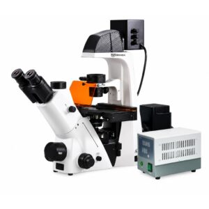
The OPXICA Inverted Phase Contrast Tissue Culture Microscope OPX-176 is designed primarily for cell culture, tissue culture and live-cell observation.
Unlike a conventional upright microscope, an inverted microscope places the objectives below the specimen. This makes it possible to observe cells and tissues directly through the bottom of culture dishes, flasks and other suitable laboratory vessels without transferring the sample to a conventional microscope slide.
The Phase Contrast system improves the visibility of transparent and low-contrast specimens without requiring routine staining. This is particularly useful when researchers need to observe living cells while maintaining their normal culture conditions.
The trinocular configuration also allows a compatible microscope camera to be connected for image capture, video recording, documentation, monitoring and digital analysis.
Applications
The OPX-176 is suitable for:
- Tissue Culture Laboratories
- Cell Culture Laboratories
- Cell Biology Research
- Biotechnology Research
- Microbiology
- Molecular Biology
- Pharmaceutical Research
- Drug Discovery Research
- Cancer Research
- Stem Cell Research
- IVF and Reproductive Biology Research
- Immunology Research
- Virology Research
- Biomedical Research
- Life Science Laboratories
- Medical Research Institutions
- Universities
- Research Institutes
- Pharmaceutical Laboratories
Typical Observation & Research Areas
The microscope can be used for observation of:
- Living cell cultures
- Tissue culture samples
- Mammalian cells
- Cell morphology
- Cell growth
- Cell attachment
- Cell proliferation
- Cell density
- Cell viability observations
- Microorganisms
- Cell culture contamination monitoring
- Drug-response studies
- Cellular morphology changes
- Biological specimens in culture vessels
Wrapping Notes
The OPXICA Inverted Phase Contrast Tissue Culture Microscope OPX-176 combines an inverted optical configuration with Bright Field and Phase Contrast observation for modern cell and tissue culture laboratories.
Its LWD Semi-APO objectives, phase contrast capability, large working stage, culture dish holder, Köhler illumination and trinocular imaging port make it suitable for routine cell observation as well as research workflows involving live and unstained specimens.
The OPX-176 is particularly suited to biotechnology, cell biology, tissue culture, pharmaceutical research, biomedical research, universities and life-science laboratories where specimens need to be observed directly within appropriate culture vessels.
Frequently Asked Questions (FAQs)
1. What is the OPX-176 used for?
The OPX-176 is primarily used for tissue culture, cell culture, live-cell observation, biotechnology research and biological laboratory applications.
2. Why is this microscope inverted?
The inverted configuration allows researchers to observe cells from below while they remain inside suitable culture dishes and vessels, making it practical for tissue-culture work.
3. Does the OPX-176 support phase contrast?
Yes. It supports Phase Contrast (PH) observation in addition to Bright Field (BF).
4. What is the standard magnification?
The specified standard magnification range is 100X–400X.
5. Which objectives are available?
The specified objectives include:
- Infinity Plan LWD Semi-APO 10X
- Infinity Plan LWD Semi-APO 20X
- Infinity Plan LWD Semi-APO 40X
- Infinity Phase Contrast LWD Semi-APO 20X
6. What type of condenser is used?
The microscope uses an LWD/ELWD condenser with N.A. 0.3 and includes a slot for a phase-contrast slide.
7. Can living cells be observed?
Yes. Phase contrast microscopy is commonly used to observe living, transparent and unstained cells, provided appropriate culture and specimen conditions are maintained.
8. Can culture dishes be placed on the stage?
The specified configuration includes a 54 mm culture dish holder and a large 210 × 241 mm working stage.
9. Can a camera be attached?
Yes. The trinocular head provides a dedicated optical path for connecting a compatible microscope camera.
10. What illumination does the OPX-176 use?
The specified transmitted illumination system uses a 6V/30W halogen light source with Köhler illumination.
11. What is the advantage of LWD objectives?
Long Working Distance objectives provide greater clearance between the objective and specimen, which can be useful when observing specimens in culture vessels or through thicker vessel bottoms.
12. Is it suitable for pharmaceutical research?
Yes. It can be used in pharmaceutical and biotechnology research for applications such as cell culture observation, drug-response studies and cellular morphology assessment, depending on the specific research protocol.