
Pathological Research Microscope: Uses, Features and Applications
Introduction
Have you ever wondered how doctors examine tiny blood cells, how scientists study human tissues, or how laboratory professionals observe microorganisms that are invisible to the naked eye?
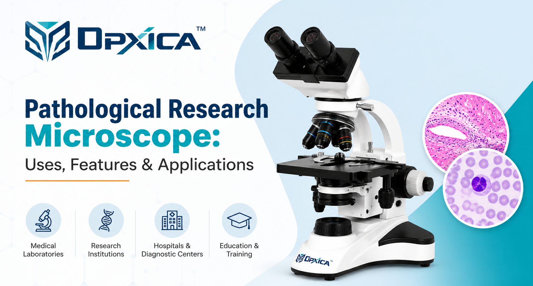
The answer often involves a powerful scientific instrument known as a Pathological Research Microscope.
A Pathological Research Microscope is an important tool in medical laboratories, hospitals, universities, and scientific research centers. It helps professionals observe microscopic structures, examine prepared biological specimens, and understand the characteristics of cells and tissues.
Modern microscopes combine advanced optical lenses, adjustable illumination, precise focusing mechanisms, and sometimes digital imaging technology to make laboratory observations clearer and more convenient.
Whether you are a medical student, laboratory technician, researcher, or scientific equipment buyer, understanding the features and applications of a Pathological Research Microscope can help you choose the right instrument.


1. What Is a Pathological Research Microscope?
A Pathological Research Microscope is a specialized compound light microscope commonly used for studying biological specimens such as tissue sections, blood smears, cells, and microorganisms.
Unlike a simple magnifying glass, this instrument uses multiple optical lenses to produce highly magnified images of small specimens.
Most conventional pathological research microscopes offer several objective lenses, commonly 4X, 10X, 40X, and 100X oil immersion. Combined with a 10X eyepiece, these provide total magnifications ranging from 40X to 1000X.
The instrument is widely used in:
-
Medical and pathology laboratories
-
Hospitals and diagnostic centers
-
Medical colleges and universities
-
Microbiology and biotechnology laboratories
-
Scientific research institutions
Advanced models may also support camera integration, digital image capture, and specialized microscopy techniques.
2. Important Parts of a Pathological Research Microscope
Understanding the main components of a microscope makes it easier to operate and maintain.
| Microscope Part | Function |
|---|---|
| Eyepiece | Allows users to view the magnified image |
| Objective lenses | Provide different levels of magnification and resolution |
| Revolving nosepiece | Holds and switches objective lenses |
| Mechanical stage | Supports and moves specimen slides |
| Condenser | Concentrates illumination on the specimen |
| Iris diaphragm | Controls the illumination aperture |
| Coarse adjustment | Helps achieve initial focus |
| Fine adjustment | Provides precise focusing |
| LED illuminator | Supplies light for observation |
| Camera port | Connects a compatible camera on supported models |
Each component contributes to the overall observation experience. Optical quality, illumination, and precise mechanical movement are particularly important for professional laboratory work.
3. How Does a Pathological Research Microscope Work?
The working principle of a Pathological Research Microscope is based on light transmission and optical magnification.
In a conventional brightfield microscope, light passes through a prepared specimen on a glass slide. The objective lens forms a magnified image, which is then viewed through the eyepiece.


The observation process is straightforward:
-
A properly prepared specimen is placed on a glass slide.
-
The slide is secured on the mechanical stage.
-
The illumination and condenser are adjusted.
-
A low-power objective is selected to locate the specimen.
-
Coarse and fine focusing controls are used to obtain a clear image.
-
Higher magnification is selected when additional detail is required.
Some advanced microscopes include digital cameras that allow users to view specimens on a computer monitor and save images for future reference.
4. Key Features of a Pathological Research Microscope
High-Resolution Optical System
The optical system determines how clearly a microscope can resolve small structures.
Research microscopes may use achromatic, plan achromatic, or infinity-corrected objectives. These optical designs help improve image quality, depending on the instrument configuration.
Multiple Magnification Levels
Different magnification levels allow users to observe specimens at different scales.
Common Magnification Combinations (Using a 10X Eyepiece)
| Objective Lens | Eyepiece Magnification | Total Magnification |
|---|---|---|
| 4X Objective | 10X | 40X |
| 10X Objective | 10X | 100X |
| 40X Objective | 10X | 400X |
| 100X Objective (Oil Immersion) | 10X | 1000X |
Formula: Total Magnification = Objective Lens Magnification × Eyepiece Magnification
Note: Higher magnification does not always mean better image quality. Optical resolution, lens quality, specimen preparation, and proper illumination also play important roles in producing clear microscopic images.
Higher magnification does not automatically mean better image quality. Optical resolution, specimen preparation, and illumination also matter.
Adjustable LED Illumination
Many modern Pathological Research Microscope models use LED illumination for consistent lighting, adjustable brightness, and energy-efficient operation.
Smooth Mechanical Stage
A mechanical stage allows controlled movement of specimen slides along the X and Y axes, making it easier to examine different regions.
Precise Focusing Controls
Coarse and fine focusing mechanisms help users locate specimens and obtain sharper images, especially when using higher magnification.
Digital Imaging Compatibility
Trinocular research microscopes can often accommodate a digital camera through an appropriate adapter.
This allows laboratories to capture photographs, document observations, and share images for teaching or research.
5. Major Uses of a Pathological Research Microscope
A Pathological Research Microscope is useful for many different laboratory activities. Its applications range from routine specimen examination to advanced biological research.

In hematology laboratories, microscopes help professionals examine red blood cells, white blood cells, and platelets in stained blood smears. These observations support the assessment of cell shape, size, and distribution.
Histopathology and Tissue Studies

Pathologists examine thin tissue sections to understand cellular arrangement and tissue structure. Stains such as hematoxylin and eosin help distinguish different microscopic components.

Laboratory researchers use microscopes to observe appropriately prepared bacteria and other microorganisms. Microscopic examination can reveal cell shapes, arrangements, and staining characteristics.
Medical colleges use research microscopes to teach histology, pathology, and cellular biology. Students learn to identify tissue structures and develop practical observation skills.
6. Applications in Different Industries
The versatility of a Pathological Research Microscope makes it valuable across multiple scientific fields.
| Industry or Institution | Main Application |
|---|---|
| Hospitals | Laboratory examination of clinical specimens |
| Diagnostic Centers | Microscopic examination of prepared samples |
| Medical Colleges | Teaching pathology and histology |
| Research Laboratories | Studying cells and tissues |
| Biotechnology Institutes | Biological sample observation |
| Pharmaceutical Research | Microscopic examination in suitable research workflows |
| Microbiology Laboratories | Observation of microorganisms |
| Universities | Practical training and academic research |
7. Types of Pathological Research Microscopes
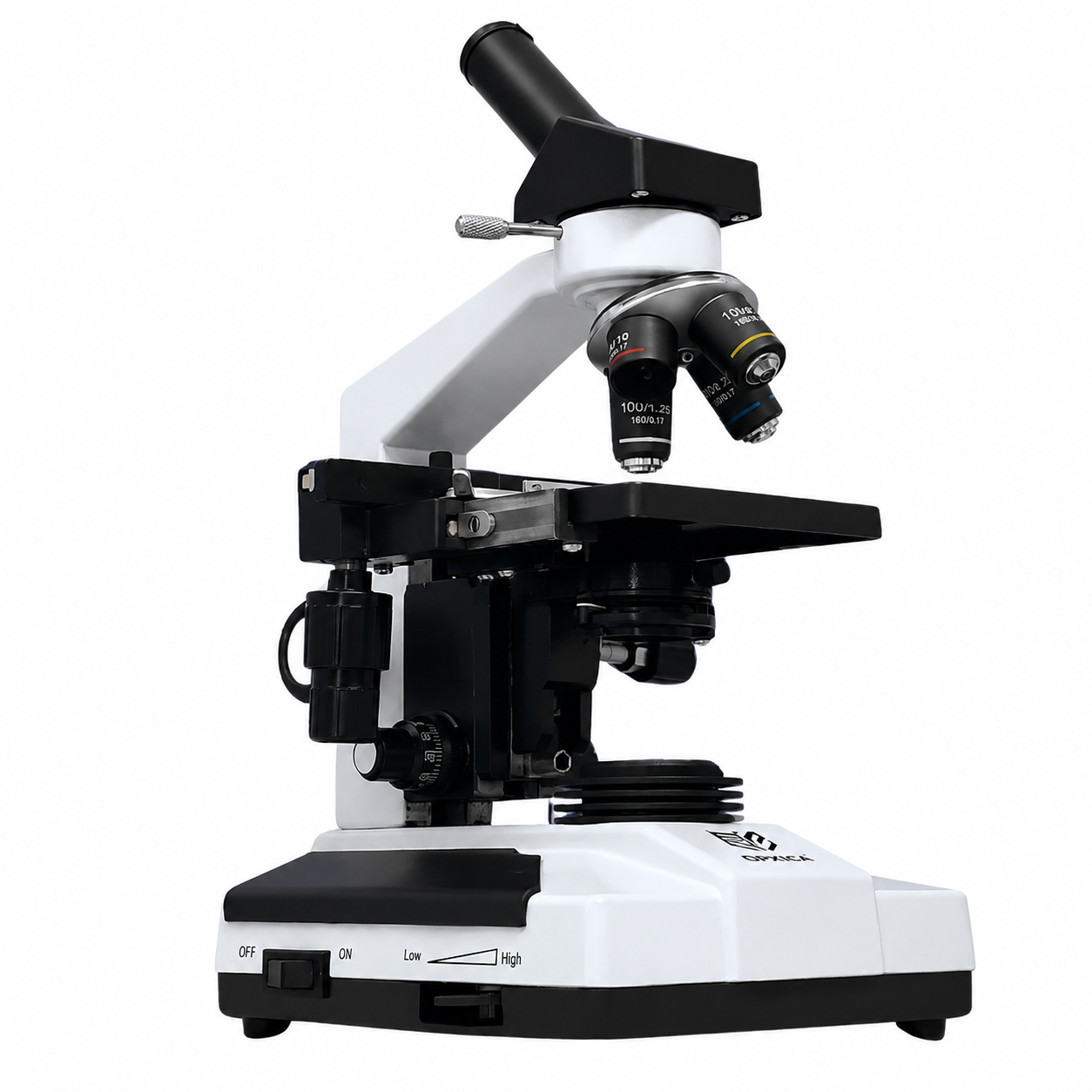
Single eyepiece, commonly used for basic teaching and observation.

Binocular Microscope
Two eyepieces for comfortable viewing during laboratory examinations.

Includes a camera port for compatible imaging and documentation.
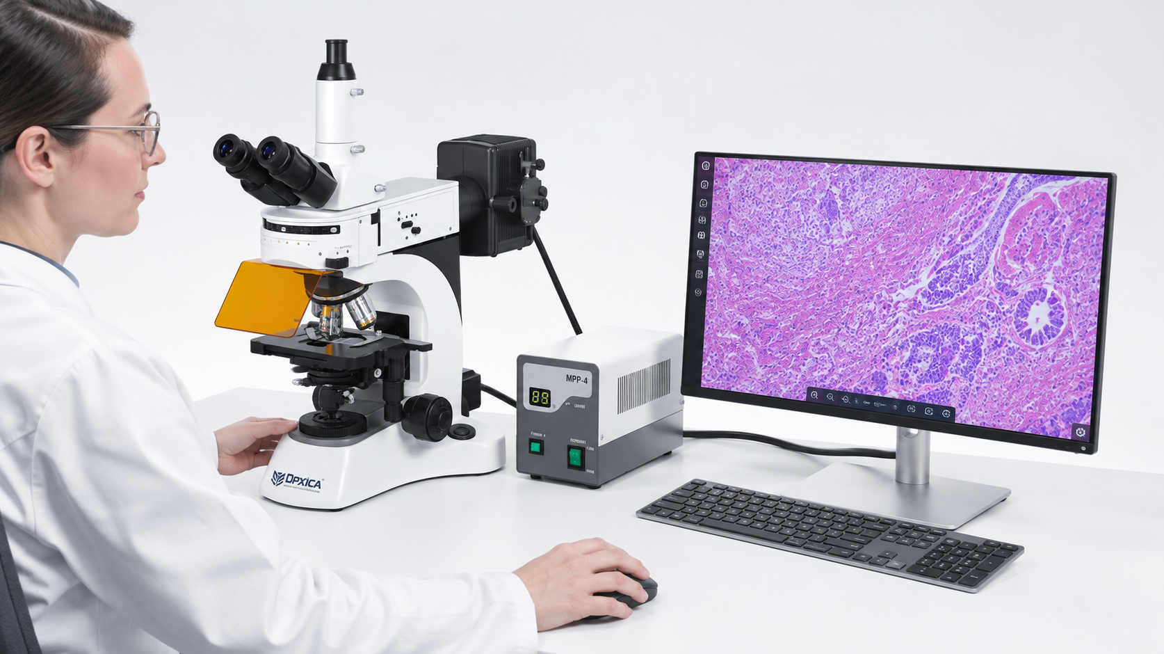
Digital Research Microscope
Supports electronic image capture, display, and documentation.
8. Pathological Microscope vs Research Microscope
Although these terms are sometimes used together, they do not always mean exactly the same thing.
| Feature | Pathological Microscope | Research Microscope |
|---|---|---|
| Main purpose | Routine pathology and specimen examination | Scientific investigation and specialized studies |
| Optical system | Commonly achromatic or plan objectives | May include advanced corrected optics |
| Camera integration | Optional | Common in advanced configurations |
| Observation techniques | Often brightfield | May support multiple techniques |
| Typical users | Pathologists and laboratory technicians | Scientists, researchers, and universities |
A high-quality microscope can serve both pathological examination and research applications, depending on its specifications.
9. How to Choose the Best Pathological Research Microscope
Choosing a microscope should begin with understanding your laboratory’s actual needs.
For routine pathological examination, optical clarity, reliable illumination, smooth focusing, and a stable mechanical stage are important.
For research applications, additional features such as infinity-corrected optics, a trinocular viewing head, camera compatibility, and specialized condensers may be beneficial.
Before selecting a Pathological Research Microscope, evaluate its optical performance, available magnifications, build quality, warranty, service support, and compatibility with your intended specimens.
A microscope with appropriate specifications is often more useful than one selected solely because of a higher advertised magnification.
10. Maintenance and Care
A properly maintained Pathological Research Microscope can deliver reliable performance over many years.
Keep the instrument covered when it is not in use, clean the lenses with appropriate optical cleaning materials, and avoid touching optical surfaces directly.
After using oil immersion objectives, clean them according to the manufacturer’s recommendations. Keep the mechanical stage free from dust and handle the focusing controls gently.
For frequently used laboratory microscopes, periodic inspection and servicing can help maintain optical and mechanical performance.
11. Frequently Asked Questions
What is the main purpose of a Pathological Research Microscope?
Its main purpose is to observe biological specimens such as cells, blood smears, tissues, and microorganisms under magnification.
What is the maximum magnification of a Pathological Research Microscope?
Many conventional models provide up to 1000X total magnification using a 100X oil immersion objective and 10X eyepiece. Specifications vary by model.
Which microscope is better for research: binocular or trinocular?
Both can provide suitable optical observation. A trinocular microscope is generally more convenient when digital camera integration and image documentation are required.
Can a Pathological Research Microscope be used in hospitals?
Yes. These microscopes are commonly used in hospital laboratories for examining prepared biological samples as part of established laboratory workflows.
Can I connect a microscope to a computer?
Yes, if the microscope supports a compatible camera system and the required imaging software.
Is a Pathological Research Microscope suitable for medical students?
Yes. It is widely used in medical education for practical training in histology, pathology, and other biological subjects.
12. Ending Notes
The Pathological Research Microscope remains one of the most valuable instruments in medical science, pathology, and biological research.
From examining stained tissue sections to studying blood cells and microorganisms, it helps laboratory professionals observe structures that cannot be seen with the naked eye.
Modern microscopes offer improved optical performance, convenient focusing, reliable illumination, and optional digital imaging capabilities.
For hospitals, universities, diagnostic centers, and research institutions, selecting the right Pathological Research Microscope can improve the convenience and consistency of microscopic observation.
The best choice is a microscope that matches the laboratory’s application, provides dependable optical performance, and is supported by appropriate maintenance and technical service.
