
Stereo Microscope: Diagram, Working Principle, Parts and Uses
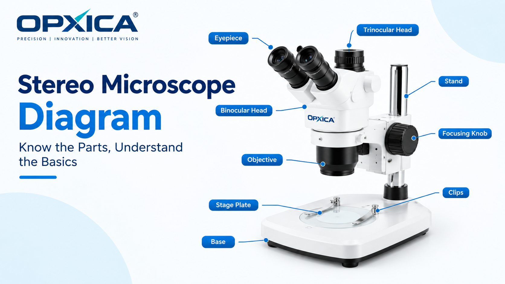
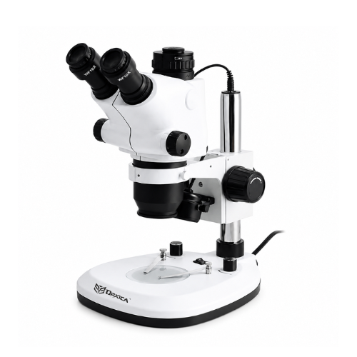
What is a stereo microscope?
A stereo microscope, also called a stereomicroscope or dissecting microscope, is a low-power optical instrument. It gives a three-dimensional, upright view of an object. It has two separate optical paths, one for each eye, and each path sees the specimen from a slightly different angle. Your brain fuses the two views into one image with real depth, the same way it does with your naked eyes.
A compound microscope looks through a thin, transparent slide at very high magnification. A stereo microscope looks at a solid object, such as an insect, a circuit board, a rock or a coin, and lets you work on it with your hands. That is why it has a long working distance, a large field of view, and an image that is not reversed or flipped.
Typical figures for a general-purpose instrument:
| Property | Typical range |
|---|---|
| Total magnification | About 5× to 100× (7×–45× is the most common) |
| Viewing angle between the two paths | About 10°–15° |
| Working distance | Several centimetres to 25 cm or more |
| Field of view | Roughly 5 mm to 50 mm across |
| Image | 3D, upright, and not laterally reversed |
| Main lighting | Reflected (incident) light, with optional transmitted light |
A short history
The idea of binocular magnification is old. The Capuchin monk Chérubin d’Orléans built a binocular instrument in 1671. It did not give a true stereo effect, because both eyes received the same image. The design that made stereoscopic microscopy practical came from the American biologist Horatio S. Greenough in the 1890s, and Carl Zeiss built it. It used two complete, separate microscopes tilted toward each other. In the 1950s, optical makers introduced the common main objective design, which now dominates research-grade instruments.
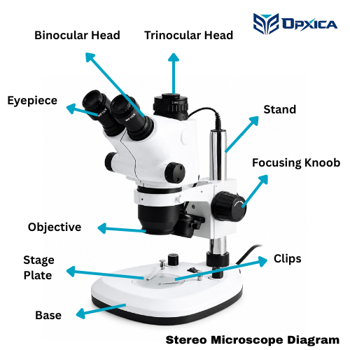
Stereo Microscope Diagram
This schematic shows the common main objective type. The light paths run from the specimen up to the eyes. The angle between the two converging paths is exaggerated so it is easy to see.
Main Parts of a Stereo Microscope
Understanding the parts of a stereo microscope makes it easier to understand its operation and applications.
1. Eyepieces
The eyepieces, or ocular lenses, are the components through which the user observes the specimen. A binocular stereo microscope has two eyepieces, one for each eye.
Eyepieces are available in different magnification powers and may include adjustable features to accommodate different users.
2. Binocular Head
The binocular head houses the two viewing channels and allows the user to observe the specimen using both eyes.
The interpupillary distance can generally be adjusted so that the two optical images merge comfortably into a single stereoscopic view.
3. Objective or Main Objective
The objective system collects light from the specimen and contributes to image magnification.
In a Greenough stereo microscope, separate objective systems are associated with the two optical paths. In a Common Main Objective system, a single large objective is shared by the two channels before the optical paths continue separately.
4. Zoom or Magnification System
Many modern stereo microscopes include a variable zoom system. The user can increase or decrease magnification while maintaining the specimen within the viewing area.
This is especially useful for inspection work because the operator can first examine a larger area at low magnification and then increase magnification to study a specific feature.
5. Focusing Mechanism
The focusing mechanism moves the microscope head or optical system relative to the specimen to obtain a sharp image.
Precise focusing is important when examining objects with uneven surfaces or different heights.
6. Stage
The stage provides a stable surface on which the specimen is placed. Depending on the microscope, it may be a simple stage, stage plate, or specialized mechanical platform.
7. Stand
The stand supports the optical head and stage and provides mechanical stability during observation.
Different stands may be designed for biological work, industrial inspection, electronics repair, or other specialized applications.
8. Illumination System
Stereo microscopes may use different illumination methods depending on the specimen.
Common arrangements include:
- Reflected or incident illumination
- Transmitted illumination
- Ring illumination
- Coaxial illumination
- Dual illumination systems
Reflected illumination is particularly useful for opaque specimens because it illuminates the surface being examined.
Stereo Microscope Principle
The stereo microscope principle is based on stereoscopic vision.
Human beings perceive depth because the two eyes observe the same object from slightly different positions. The brain processes these two images and interprets their differences as depth.
A stereo microscope uses the same basic concept.
Instead of sending exactly the same image to both eyes, the microscope provides slightly different optical views of the specimen to the left and right eyes. The brain combines these views, producing a three-dimensional visual impression.
In simple terms:
Two optical views → Two eyes → Brain combines the views → Perception of depth
This is the fundamental stereo microscope principle.
The two optical paths are arranged so that each eye receives a view from a slightly different angle. This provides stereoscopic depth information that is particularly valuable when examining three-dimensional objects or manipulating specimens under magnification.
How Does a Stereo Microscope Work?
The working process of a stereo microscope can be explained in several stages.
Step 1: Illumination of the Specimen
Light is directed toward the specimen. For many opaque objects, illumination comes from above or from another direction so that light reflects from the surface.
Step 2: Collection of Light
The objective system collects light reflected from the specimen.
Step 3: Formation of Two Optical Views
The microscope creates two optical paths corresponding to the left and right eyes.
Depending on the microscope design, this may occur through two separate objective systems or through a common main objective followed by two separate optical channels.
Step 4: Magnification
The optical system magnifies the specimen. In a zoom stereo microscope, magnification can be continuously adjusted across the available zoom range.
Step 5: Image Delivery to the Eyes
The left and right optical channels deliver slightly different views to their respective eyepieces.
Step 6: Stereoscopic Perception
The user’s brain combines the two images and produces the perception of depth.
This allows the operator to see not only the surface details of the specimen but also its three-dimensional form.
Optical Designs of Stereo Microscopes
There are two major optical arrangements used in stereo microscopy.
1. Greenough Stereo Microscope
The Greenough design uses two separate optical systems, one for each eye. The optical axes are arranged at an angle so that the left and right eyes receive different perspectives of the specimen.
Advantages
- Compact optical construction
- Effective stereoscopic viewing
- Suitable for routine inspection and educational applications
- Often available in relatively simple configurations
The Greenough concept has been used in stereo microscopes for more than a century and remains an important optical configuration.
2. Common Main Objective (CMO) Stereo Microscope
In a Common Main Objective (CMO) design, both viewing channels share a common main objective at the front of the microscope. Behind the objective, the optical paths are separated into two channels.
CMO systems are often used in more advanced stereo microscopes because their architecture can accommodate various optical accessories and imaging configurations.
What is a binocular stereo microscope?
A binocular stereo microscope is a stereo microscope designed for observation using both eyes.
The term “binocular” simply indicates that there are two viewing channels. However, it is important to understand that not every binocular microscope automatically provides true stereoscopic vision.
A conventional compound microscope may have a binocular viewing head that splits one image into two eyepieces. In that arrangement, both eyes may essentially see the same image.
A true stereo microscope provides separate optical views to the two eyes, allowing the brain to generate stereoscopic depth perception.
Stereo Microscope Uses
The stereo microscope uses are broad because the instrument is particularly useful for objects that need to be viewed as three-dimensional structures.
1. Biological Dissection
Stereo microscopes are widely used for dissection and manipulation of biological specimens.
They provide enough working distance for instruments such as forceps, needles, scalpels, and probes to be used while observing the specimen.
2. Entomology
In entomology, stereo microscopes are used to examine insects and other small organisms.
Researchers can study:
- Body structures
- Wings
- Legs
- Antennae
- Surface patterns
- Small morphological features
The three-dimensional view can make manipulation and orientation of specimens easier.
3. Botany and Plant Studies
Stereo microscopes can be used to observe plant structures such as:
- Seeds
- Leaves
- Flowers
- Buds
- Roots
- Small plant parts
They are particularly useful when the specimen is too large or three-dimensional for convenient examination under a compound microscope.
4. Electronics and PCB Inspection
One of the important industrial stereo microscope uses is inspection of electronic components and printed circuit boards.
Operators can examine:
- Solder joints
- PCB tracks
- Connectors
- Small components
- Surface defects
- Assembly quality
The combination of magnification, depth perception, and working distance makes stereo microscopes useful for inspection and rework operations. Government laboratory C-MET, for example, lists PCB and substrate inspection among stereo microscope applications.
5. Jewelry and Gemstone Examination
Jewelry manufacturers and gemologists can use stereo microscopes to inspect stones, settings, surface features, inclusions, and workmanship.
The stereoscopic image helps users understand the shape and spatial position of small features.
6. Watchmaking and Precision Assembly
The large working distance of stereo microscopes allows small mechanical parts to be manipulated while being observed under magnification.
This makes them useful in watchmaking, miniature assembly, repair, and other precision tasks.
7. Industrial Inspection
Stereo microscopes are widely used in manufacturing and quality control.
They can assist in examining:
- Surface defects
- Small mechanical components
- Plastic parts
- Metal components
- Machined surfaces
- Assembly quality
- Fracture surfaces
Stereo microscopy is particularly useful when an operator needs to inspect a physical object rather than a thin prepared slide.
8. Forensic Examination
Stereo microscopes can be used for preliminary examination and comparison of physical evidence.
Applications may include examination of:
- Fibers
- Insects
- Small fragments
- Tool marks
- Surface features
- Material evidence
9. Geology and Mineralogy
Rocks, minerals, crystals, and geological samples often have complex three-dimensional surfaces.
Stereo microscopy can help researchers examine:
- Crystal structures
- Grain characteristics
- Surface texture
- Mineral fragments
- Small geological specimens
10. Educational Laboratories
Stereo microscopes are useful in school, college, and university laboratories for demonstrating three-dimensional specimens.
Students can observe insects, plant structures, seeds, rocks, small mechanical components, and other specimens without requiring complex slide preparation.
Why is Working Distance Important in a Stereo Microscope?
One of the major advantages of a stereo microscope is its relatively large working distance.
Working distance refers to the distance between the front of the objective system and the specimen when the specimen is in focus.
A useful working distance provides space between the lens and the object, allowing the user to position tools or manipulate the specimen.
This is especially valuable for:
- Dissection
- Electronics repair
- Precision assembly
- Jewelry work
- Industrial inspection
- Biological specimen preparation
Working distance and depth of field are important characteristics of stereo microscopy because the instrument is intended not only for viewing but also for practical manipulation of three-dimensional specimens.
Stereo Microscope vs Compound Microscope
| Feature | Stereo Microscope | Compound Microscope |
|---|---|---|
| Viewing | Three-dimensional/stereoscopic | Generally flat two-dimensional image |
| Optical paths | Two separate viewing paths | Usually one primary optical path |
| Typical application | Solid objects and surface inspection | Thin specimens and microscopic structures |
| Magnification | Low to moderate | Moderate to very high |
| Working distance | Generally larger | Generally shorter |
| Specimen manipulation | Easy during observation | More restricted |
| Illumination | Frequently reflected light | Commonly transmitted light |
| Depth perception | Major feature | Not true stereoscopic depth perception |
| Common uses | Dissection, electronics, insects, jewelry, inspection | Cells, tissues, microorganisms, prepared slides |
The distinction is not simply about magnification. The optical design and intended specimen type are equally important. Stereo microscopes prioritize depth, working distance, and specimen handling, whereas compound microscopes are generally optimized for high-resolution observation of microscopic structures.
Advantages of a Stereo Microscope
Stereo microscopes offer several practical advantages:
1. Three-Dimensional Observation
The two optical channels provide stereoscopic depth perception.
2. Large Working Distance
Users can manipulate specimens while observing them under magnification.
3. Wide Field of View
Lower magnification allows the operator to see a relatively large area of the specimen.
4. Easy Specimen Handling
Objects can often be examined directly without preparing thin microscope slides.
5. Comfortable Binocular Observation
Using both eyes can make prolonged inspection more comfortable than monocular observation.
6. Suitable for Solid Objects
Three-dimensional and opaque specimens can be examined effectively.
7. Flexible Magnification
Zoom stereo microscopes allow users to move between overview and detailed inspection without changing the specimen.
Limitations of Stereo Microscopes
Stereo microscopes are not intended to replace high-power compound microscopes in every application.
Their limitations include:
- Lower maximum useful magnification than many compound microscope systems
- Lower resolution for very fine cellular structures
- Not primarily designed for viewing thin biological sections
- Optical performance depends strongly on objective, eyepiece, illumination, and microscope design
- Increasing magnification can reduce field of view and working distance
Therefore, the correct microscope depends on the specimen and the type of observation required.
How to Choose a Stereo Microscope?
When selecting a stereo microscope, several specifications should be considered.
Magnification Range
Select a magnification range that matches the smallest details that need to be observed.
Working Distance
A larger working distance is useful when tools must be used under the microscope.
Optical System
Consider whether a Greenough or Common Main Objective system is appropriate for the intended application.
Zoom Ratio
A larger zoom range provides greater flexibility between overview and detailed inspection.
Illumination
Select reflected, transmitted, coaxial, ring, or combined illumination according to the specimen.
Binocular or Trinocular Configuration
A binocular system is suitable for visual observation. A trinocular configuration may be preferable when a camera needs to be integrated for documentation or image analysis.
Ergonomics
For long inspection sessions, consider the viewing angle, interpupillary adjustment, focusing controls, stand design, and operator comfort.
Frequently Asked Questions About Stereo Microscopes
What is a stereo microscope?
A stereo microscope is an optical microscope that provides a three-dimensional view of a specimen by delivering slightly different views to the left and right eyes through separate optical paths.
What is the principle of a stereo microscope?
The stereo microscope principle is based on stereoscopic vision. Two slightly different optical views are delivered to the two eyes, and the brain combines them to create the perception of depth.
What is a stereo microscope used for?
Common stereo microscope uses include biological dissection, entomology, electronics and PCB inspection, jewelry examination, watchmaking, industrial quality control, forensic examination, geology, and educational laboratory work.
What is a binocular stereo microscope?
A binocular stereo microscope is a stereomicroscope that provides two viewing channels for observation with both eyes. In a true stereo configuration, the two channels provide different perspectives that create stereoscopic depth perception.
What is the difference between a stereo microscope and a compound microscope?
A stereo microscope is designed primarily for three-dimensional observation and manipulation of solid specimens at relatively low to moderate magnification. A compound microscope is generally designed for higher-magnification examination of microscopic structures, often using prepared slides.
Why does a stereo microscope produce a 3D image?
A stereo microscope provides slightly different views of the specimen to the left and right eyes. The brain combines these views in a process called stereopsis, producing the perception of depth.
Can a stereo microscope be used for electronics?
Yes. Stereo microscopes are commonly used for PCB inspection, solder-joint examination, component inspection, repair, and precision electronics work.
At the End
A stereo microscope is designed for situations where depth perception, surface detail, working distance, and specimen manipulation are more important than extremely high magnification. Its two optical viewing paths provide different perspectives of the specimen, allowing the brain to create a three-dimensional visual impression.
From biological dissection and entomology to PCB inspection, jewelry, geology, manufacturing, and education, the stereo microscope uses cover a wide range of scientific and industrial applications.
Understanding the stereo microscope principle, optical designs, working distance, illumination, magnification, and binocular viewing system can help laboratories, researchers, students, and industrial professionals select the right microscope for their specific requirements.
Whether you are looking for a microscope for biological dissection, industrial inspection, electronics work, or educational applications, a properly configured binocular stereo microscope can provide a practical combination of magnification, depth perception, and specimen accessibility.
